Digital Radiography
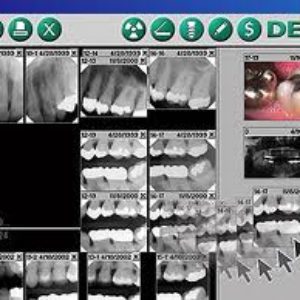
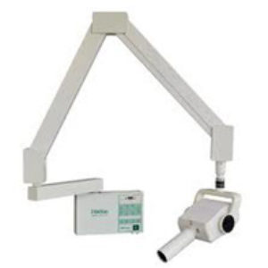
The physical process for digital radiography is actually similar to traditional dental X-rays that use film: With digital radiography, your dentist inserts a sensor into your mouth to capture images of your teeth -- but that's where the similarities between conventional and digital dental X-rays end. Although it resembles the film used for bitewings and other X-rays, the digital sensor is electronic and connected to a computer. Once the X-ray is taken, the image is projected on a screen for your dentist to view.
There are several benefits to using digital radiography over traditional film X-rays:
Less Radiation -- The equipment used in digital radiography exposes dental patients to much less radiation. In fact, digital X-rays use up to 90 percent less radiation than film X-rays. While conventional dental X-rays are relatively safe, digital radiography is an excellent option for those who take X-rays on a regular basis or for those who are concerned about radiation.


Dental X-rays are an important part of the dental exam, used to diagnose problems with the teeth, gums and jaw. X-rays use electromagnetic radiation to produce these images -- they form wavelengths that penetrate the soft tissue of the body and are absorbed by denser materials, creating pictures of your bones or teeth. And since your dentist doesn't have X-ray vision, they're necessary to distinguish dental problems not visible to the naked eye. Tooth decay, periodontal disease, impacted teeth, bite problems and even tumors are just a few of the dental conditions easily found with dental X-rays.
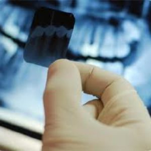
Bitewings are a type of dental X-rays used to check the back teeth. As they show the crown of the tooth, bitewing X-rays are extremely helpful in determining tooth decay located in between teeth, various stages of gum disease and problems with tooth alignment. Bitewings are also excellent for detecting a buildup of dental tartar, and are sometimes used to measure bone loss due to advanced periodontal disease.
Spreading Their Wings
Bitewings get their name from how they look: The film resembles a "T-shape" that's placed on the interior side of the jaw. The film extends to cover both the upper and lower teeth, and the patient bites a tab in the middle to hold the film in place. After our dental assistant positions the bitewing in the mouth, the patient closes his or her teeth to secure the film. An X-ray camera is then used to photograph several teeth at once
Woodland Hills
Topanga
Malibu
Canoga Park
Tarzana
West Hills
Winnetka
Hidden Hills
Calabasas
Granada Hills
Arleta
Santa Clarita
Northridge
Encino
Sherman Oaks
Panorama City
Van Nuys
Simi Valley
Reseda
Lake Balboa
Westlake Village
Hidden Valley
Saratoga Hills
Newbury Park
Oxnard
Camarillo
Agoura Hills
Oak Park
Thousand Oaks
Moorpark
Porter Ranch
Granada Hills
North Hills
Studio City
Los Angeles
Ventura
Bell Canyon
Hidden Valley
Saratoga Hills


